My Microbe Academia
This section has now been turned into a place where I could brain dump all of my notes (Reminder: I am in my last year in Microbiology undergrad). It have been unfortunately reminded that I am quite weak in the executive functioning department, and so I must work with some kind of accountability in order to actually do my shit. I know nobody reads this blog anyway, so I might as well, right? Note that this is written with the idea that I will be the one rereading. That's all!
An important note: There are likely typos here! I come back to it a lot to review/proofread but I could still miss stuff. Thanks for understanding! Also also! If you click the pictures, it's going to take you to the sites where I took them, and could give you more insight on the subject.
[4 Y 1 S] M I D T E R M S
[MMC] Medical Microbiology Lecture
1. Introduction to Medical Microbiology
Date Finished: October XX, 2024
We all know that diseases come from microorganisms. "Germs", one would say. Which is why Micrbiology and Medicine has always been connected. Medical Microbiology is their baby. It's studying microorganisms with a focus on what they do to our health, and how to treat them.
Be alert, for this lesson is filled with definitions and explanations. I will try to be as clear and organized as possible here. Let's go!
Medical Microbiology is the study of pathogens, the diseases they cause, and how our body tries (and fails sometimes) to protect us from them. It is concerned with prevention, diagnosis and treatment of infectious diseases.
Clinical Microbiology, also known as Diagnostic Microbiology, is only a branch of MM that is only concerned with the the study of the pathogens and their roles for disease. This is basically what happens when the patients give sampoles to the hospital laboratory and they culture that in order to find out what naughty microbe is hurting them, and make antibiotic susceptibility tests to see what antibiotics will work against them. Remember when you did OJT? The lab results? That.
Of course an introduction to a branch of micrbiology won't be complete without a little history! We only got four this time, but they're pretty important.
| Name | Important Thing | Context |
|---|---|---|
| Edward Jenner | Vaccine | Basically he looked at cowpox and went, 'Hmm... that looks a lot like smallpox. And cows who have cowpox no longer gets it again, so maybe we can just get cowpox pus from this lovely cow and inject this into this lovely little boy and see what happens!' I feel bad for the boy because he was literally exprimented on but... science? Vacca means cow, btw. The more you know. |
| Louis Pasteur | Pasterurization; Swan Flask Experiment; vaccines against anthrax, fowl cholera, and rabies | Basically this dude looked at the theory of spontaneous generation, decided that's not right and decided to make an experiment (picture below). Also lots of other stuff. He's an accomplished and busy man. |
| Robert Koch | Koch's Postulate | He and Pasteur are tw of the main founders of modern bacteriology. He also helped connect a lot of diseases with the specific microbes that caused them through the Koch's postulates. |
| Joseph Lister | Antiseptic surgery | Back then, surgeons don't sterilize their tools during surgery so a lot of people die. He was the first to try to do so using carbolic acid solution and also used it to clean ipen wounds after surgery. |
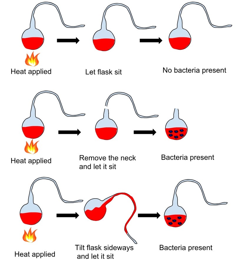
This is Pasteur's Swan Neck Flask Experiment, which disproved spontaneous generation. Back then, people thought that food spoils because maggots, molds, etc., spontaneously generate on the food and that causes it to spoil. But he proved that the broth inside the flask won't go bad as long as there is no bacteria present in it.
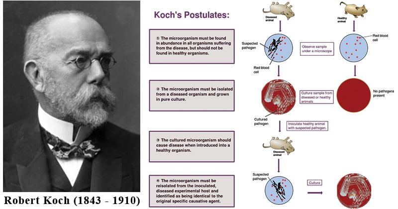
This is Koch's Postulates, which he used to figure out what bacteria causes which disease. But it has a lot of limitations.
Limitations of Koch's postulates:
- Not all pathogens can be cultured, like cerain viruses and bacteria who are fastidious.
- Some carriers of pathogens are asymptomatic
- Many diseases are polymicrobial, or caused by many different pathogens.
- Thing that are applied to animal models may not accurately apply to humans
- Host may respond differently to the same pathogen, depending on genetics and immune responses.
There are many fields under Medical Microbiology. It includes microbial physiology, microbial genetics, parasitology, virology, immunology and serology(antibodies). All these branches work together to better understand and treat human diseases.
The human body always has microbes on or in it. Always. That is why there is something that is called natural microflora, which are the normal microbiota present in our body. These microbes make up 10x the total number of cells in our body. Microbes are commonly present in our skin, mouth, throat (nasopharynx and oropharynx), gastrointestinal tract, and genitourinal track. If they get into your blood, lymph, spinal fluid or internal tissue and organs (which are supposed to be sterile), that indicates an infection.
A newly born baby has no natural microflora yet. They are sterile during pregnancy. Or at least, they're supposed to be. Sometimes expecting mothers pass on viruses to their babies, but that's another thing entirely. Babies get their microflora normally during childbirth, when they get in contact with their mother's birth canal. They also get even more microbiota from their environment.
In contrast to our natural microflora, there is the transient microflora, which are the microbes that are only temporary in our body. If they are acapable of causing disease, they are considered true pathogens. Examples are the Influenza virus, plague bacillus, and malarial protozoans. They often attach but cannot proliferate because our natural microflora protects us through microbial antagonism. Basically they out-compete the newbies and drive them off. This is why disruption of natural microflora may lead to disease, like how prolonged use of antibiotics may lead to diarrehea because our gut microbes die off, allowing bad bacteria to prolierate in our intestines.
But that doesn't mean our natural microflora is all goody-goody. Sometimes they can be Slytherin too. Opportunistic pathogens are organisms that are part of our microflora, but when there is an opportunity, like weak immune system or changes in the environment, they proliferate, causing diease. An example is Candida albicans, which is a yeast that grows in the vagina. When the pH becomes basic, they grow too fast and cause Candidiasis (aka yeast infection). Another example are Pseudomonas spp., which can cause pneumonia, urinary tract infection, etc.
Sometimes taking advantage of this concept can actually lead to better health, such as in the case of biotherapeutic agents, which involves reinoculation of the human body by beneficial microbes. An example is drinking probiotics (probiotics means food with live microorganisms, prebiotics mean food that has nutrients to allow them to grow better. Some food has both) to get some good ole gut bacteria, or in some extreme cases: there is a procedure called fecal transplant, where a doctor literally takes poop from a relative or housemate and shoves it in your large intestine. Sometimes, you just gotta do what you have to do, I guess.
Epidemiology and Public Health
Naturally, Medical Microbiology is involved with these two subjects. But let's define them first.
Epidemiology is a science that involves studing the incidence and distribution of diseases in big populations,and trying to figure out what could have caused the spread, and how sever these diseases are. Basically, they are the study of the 'epidemicity' (is that a word?) of diseases. How widespread is it? Why is it spreading? Yanno?
Public Health, on the other hand is concerned with protecting the health of entire populations through community-wide action. This is very intertwined with government agencies and the projects they make to help keep people healthy. This is concerned with the ENTIRE population, not just one person.
More definitions! Hoooray (sarcasm).
Statistics of Disease
- Prevalence - How much of the population is sick of this disease? This is presented with a percentage.
- Incidence - How many new cases are there over a certain time period? (For example, in a month, how many new patients got sick?)
- Herd Immunity - This is something that happens when most of the population is immune, which typically happens when the are vaccinated. There is some sort of computation for this, but supposedly, when you reach a certain point, the herd would have immunity against the disease/
- Morbidity rate - How many are sick?
- Mortality rate - How many died?
Scale of Disease
- Sporadic - shows up occassionally at irregular intervals
- Endemic - relatively steady frequency over a long period of time in a specific area
- Hyperendemic - the endemic disease has increased but its not an epidemic yet. Must keep tabs on it.
- Epidemic - rate of disease is increasing beyond expected. This is now an outbreak.
- Pandemic - Epidemic across continents. Like COVID-19.
Microbe-Human Interactions
- Infection - If a microbe went inside you and started to multiply already, you are infected. The bacteria are the colonizers and your body is the New World or something, idk
- Disease - dirsuption of tissue or organs caused by microbes and their productsl; basically anytime that you're not healthy.
- Pathology - study of disease, including cause (etiology), step by step progression (pathogenesis), and effect on the normal structure and functioning of the body.
- Host - organism that shelters and supports the pathogens. If you're sick, it's YOU.
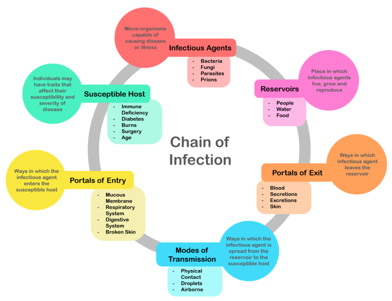
This is the Infectious Disease Cycle. Basically it starts with the Host. When an infectious agent comes in, the host becomes a reservoir. After the agent proliferates inside the host, it exits the host to infect more people. Through different modes of transmission, it finds a new portal or entry for a new host, and the cycle starts again.
[MEC] Microbial Ecology Laboratory
1. Microscopy
Ah, microscopy. The topic we come back to and re-discuss at least twice at the beginning of each semester. I suppose as tools of the trade, it just makes sense the professors want us to understand it forwards, backwards, and upside down. So here we go!
There are many types of microscope, but there are the main two: Light Microscope, which uses light to magnifiy objects, and electron microscope, which uses electrons. For the purposes of this lesson, let us stick to the light mircroscope.
Compound Light Microscopes are microscope that uses multiple lenses in order to magnify objects. It has three parameters: Magnification (how big?), Resolution (how clear? how distinguishable?) and Contrast (dark vs light, foreground vs background).

Behold! The microscope.
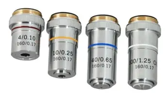
There are four types of objectives on a common compound light microscope, and they have a corresponding color coding as seen here. They are the Scanner (4x; red), Low Power Objective (10x; yellow), High Power Objective (40x; blue) and Oil Immersion Obective (100x; white).

The eyepiece would typically have these numbers on the side. The 10x indicate the magnification of the ocular lens, while the 23 indicates the field number, which corresponds to the field of view, or how big of a space is the area that can be seen under the microscope.
A couple of terms to remember:
- Focal Length - How far away is the observer from the slide?
- Working Distance - How far away is the objective from the slide?
- Resolving Power - How clear is the resolution? (Is it HD)
- Numerical Aperture - How wide is the "eye" of the objective? How good is it at collecting light?
- Parfocus - ability to keep focus after objective change.
- Stage Aperture - part of the microscope. Like numerical aperture, but for the stage.
- Diaphragm - part of the microscope located under the stage. Acts like a curtain to control light.
- Condenser - part of the microscope located under the stage. Gathers light and focuses it on the specimen.
[MEC] Microbial Ecology Lecture
2. Diversity of Microorganisms
Date Finished: October XX, 2024
Microorganisms are all around us in the environment. Some can even thrive in extreme environments. Like:
| hyperthermophiles | >121 deg Celcius |
|---|---|
| psychrophiles | <20 deg Celcius |
| halophiles | salterns, Dead Sea |
| acidophiles | acid mine drainage |
| alkaliphiles | playa lakes |
Note: Most extremophiles are Archaea, but not all Archaea are extremophiles. Likewise, there are some extremophiles from Bacteria as well. (Nature, unfortunately, is chaotic and does not surrender to the order humans wanted to limit it to.)
The morphologies of bacteria and achaea is diverse. They modify shape for adaptation.
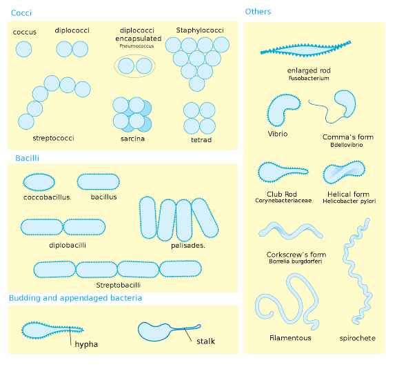
This is a picture of different morphologies. It is not all of them, but these are the most common. There is another type, called sprilla. It is similar to spirochete, but spirochete is more flexible.

Certain species of Caulobacter uses an appendage called a prothescate which is important for them to attach to surfaces.
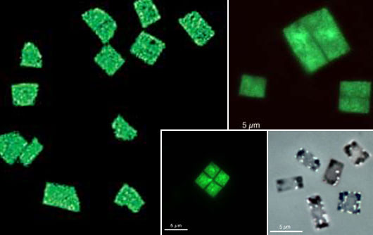
There is a square shaped bacteria! It's called Haloquadratum walsbyi. The name comes from salt (halo), square (quadratum), and the name of the person who discovered it, John Walbsy (walsbyi). I wonder what forced them to evolve this way? Note to self: research later.
Three Domains of Life
These are the Three Domains of Life. According to Carl Woese in his study of 16S RNA, Archaea is actually closer to Eukarya than Bacteria. LUCA means Last Universal Common Ancestor.
Similarities and Differences Between the Three Domains:
- Cell Wall
Eukarya has no peptidoglycan.
Archaea and Bacteria cell wall structure is a bit more complicated.
All of the domains sometimes have cells that has no cell walls. - Cytoplasm Membrane
Eukarya and Bacteria has glycerol esters of fatty acids.
Archaea has glyerol ethers of fatty acids. Remember Ethan (my OC)? His species is "archaic", so Ether. - Genetic Material
Eukarya and Archea both have histones.
Archaea and Bacteria both have circular chromosomes and plasmids.
Eukarya has linear chromosomes and a nucleus. - RNA polymerases
Eukarya has 3 RNA pol (12-14 subunits)
RNA pol I codes ribosomal RNA RNA pol II codes messenger RNA RNA pol III codes transfer RNA *(RMT... Resident Medical Technologist??? HA)
Archaea has 1 RNA pol (8-12 subunits
Bacteria has 1 RNA pol (4 subunits) - Transcription Factors and Antibiotic Resistance
Eukarya and Archaea require transcription factors, Bacteria does NOT. (Transcription factors are proteins that help turn specific genes "on" or "off" by binding to nearby DNA.)
Bacteria is the only one affected by most antibiotics. (I say most, because you never know...)
Bacteria Cell Wall Structure
There are mainly two types of bacterial cell wall. There is the Gram-positive and the Gram-negative.
The cell wall of bacteria contain peptidoglycan aka murein. This peptidoglycan is composed of sugar and peptide chains. The sugar chains, in this case, are N-acetylglucosamine and N-acetylmuramic acid. Note those down. They are important.

Gram-positve vs Gram-negative cell wall Structure. Gram-postive has a thick peptidogylcan layer while Gram-negative has a thin one.
Gram-positive vs Gram-negative Bacteria Face-off (In Layers)
Note: Remember that nature does not conform and that some of these structures may be absent/different in some bacteria.
| GRAM-POSITIVE | GRAM-NEGATIVE | NOTES |
|---|---|---|
| OUTSIDE OF THE CELL | ||
| teichoic acid1 | lipopolysaccharide (LPS)2 | Both are anchored on the surface (1) for cell wall integrity, ion transport, and adhesion (2) for cell wall intergrity, antibiotic/detergent resistance, and adhesion. Can trigger strong immune response. |
| peptidoglycan3 + lipotechoic acid4 | outer membrane8 with porin (hole) | (3) this layer is THICC (4) anchors the peptidoglycan layer to the cell membrane. Also for cell integrity, adhesion and ion regulation. Can trigger immune responses. |
| periplasmic space5 + lipoprotein6 | (5) small gap between membrane and peptidoglycan wall (6) anchors outer membrane to pepti layer and aids in transport, enzymatic processes and bacterial response to environment (biofilm formation) | |
| peptidogylcan7 | (7) this layer is THIN | |
| periplasmic space (again) | ||
| plasma/cell membrane8 | contains membrane proteins and phospholipis | |
| HOORAY! INTERIOR OF THE CELL! | ||
But what about Archaea?
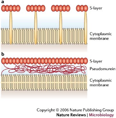
The structure of Archaeal cell wall.
Archea don't have peptidoglycan. They have pseudopeptidoglycan also known as pseudomurein. It consists of N-acetylglucosamine (same as bacteria!) and N-acetyltalosaminuronic acid, which is NOT the same as in bacteria. Some archaea however, don't have pseudomurein, but instead have cell walls made of other, less specialized polysaccharides.

There is a structure common to Archaea which can also be found in some bacteria, called the S-layer, which is a layer of proteins/glycoprotein subunits which forms a crytalline lattice for protection of the cell.
Whew! That's a lot. Okay, let's move on.
Let's move on to Diversity!
Microorganisms just in the soil is diverse. Through an experiment by Sergei Winogradsky called the Winogradsky column, this can be demonstrated. He studed chemolithotrophy (ability to obtain energy by using inorganic compounts) and chemoautotrophy (using that energy to convert CO2 to produce their own organic matter.
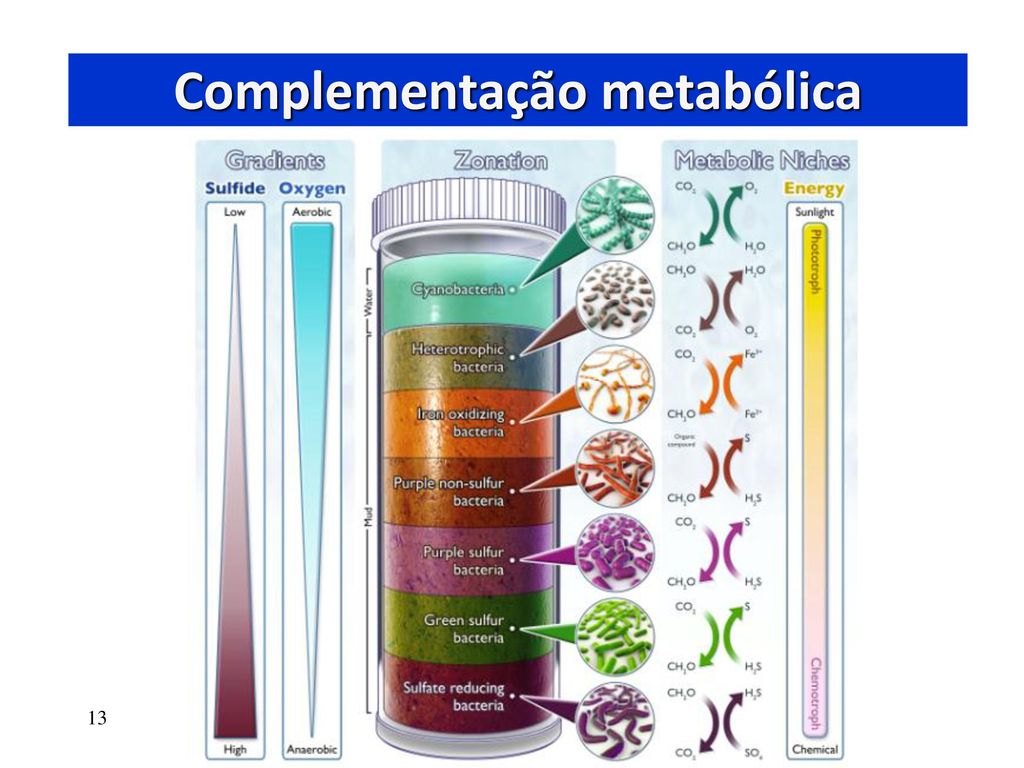
The Winogradsky Column. As you can see there are many layers to it. Remember to memorize them all! But why are the bacteria in the column where they are?
| Bacteria | ,Reason |
|---|---|
| Cyanobacteria | They are photosynthetic, and thus need the sun. So they are at the top. They are primarily aquatic, too. |
| Heterotrophic bacteria | They use organic matter (dead plants and animals) for energy, so they need to be close to surface where there are more of those. Also, most of them are aerobic (though some aren't). |
| Iron-oxidizing bacteria | Most of them are aerobes. However, concentrations of iron is higher deeper underground. So they compensate by staying arounf the middle. |
| Purple non-sulfur bacteria (PNSB) | They are versatile and can photosynthesize but also use organic compounds for energy. So they linger closer to the surface. However, most of them are facultative anaerobes, which means despite being able to live in oxygen, they prefer without. So they're a bit deeper in the column. |
| Purple sulfur bacteria (PSB) | They are deeper than their PNSB brothers because they want more sulfur. And sufur concentrations are higher down there. |
| Green sulfur bacteria (GSB) | They are able to photosynthesize in deeper light, so they can be close to the bottom. They want sulfur too, obviously. |
| Sulfate-reducing bacteria | Decomposition of organic matter above can lead to accumulation of sulfates deeper down. They love it. |
Diversity of Bacteria
Using small subunit ribosomal RNA, we have recognized and noted 89 bacterial phyla, but true count can reach up to 1500. (That's a lot!)
Over the years as we discover more of these species, the number of phyla increased:
| Year | Scientist | No. of phyla |
|---|---|---|
| 1987 | Woese | 11 |
| 2002 | Hugenholtz | 36 |
| 2003 | Rappe and Giovannoni | 52 |
| 2016 | Solden et al | 89 |
So what are some groups of bacteria? (This are simply examples of their diversity and is by no means an exhaustive list)
- Phototrophs
These are bacteria that get energy from the sun. They use their photochemical reaction centers (a fancy way to say proteins that activate with sunlight) to do that through electron transfer reacions.
Phyla:
- Chlorobi


Chlorobi are green-sulfur bacteria. They are actually green because they have bacteriochlorophyll a! The green stuff you see in stagnant ponds has green-sulfur bacteria.
Notable information:
- They are obligate anaerobe, and thus don't produce oxygen.
- They oxidize (take electrons from) sulfur
- They reduce (add electrons, damn you old scientists for making this so confusing) iron or hydrogen.
- They have Photosystem I.
- They have a special light-harvesting protein called the Fenna-Matthews-Olson (FMO) Complex.
- Cyanobacteria
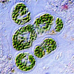
Cyanobacteria are the typical oxygen-makers in the bacterial world. They make 20-30% of the world's oxygen. They are also called blue-green algae, but that is a mismoner because they are definitely not true algae.
Notable information:
- They have both Photosystems I and II.
- They use Calvin Cycle to reduce CO2, like plants
- They have sepcial light-harvesting antennae called phycobilisomes, which can also be present in some red algae.


The structure and position of a phycobilisome.

To put things into perspective, this is what a cyanobacteria structure looks like. As you can see, there are thylakoid membranes in the cytoplasm, and the phycobilisomes are embedded there.
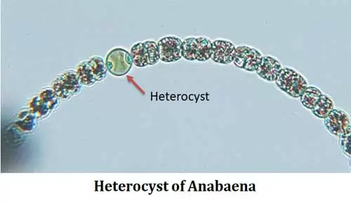
On the other hand, another interesting thing that cyanobacteria do is forming heterocysts. These are anaerobic containers that are as big or sometimes even bigger than the cells themselves, which is created for nitrogen fixation because the enyzyme responsible for it, Nitrogenase, is sensitive to oxygen.
- Chloroflexi
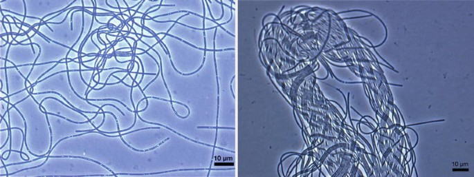
Chloroflexi are filamentous bacteria. They look like forbidden spaghetti noodles!
Notable information:
- They are gliding bacteria. Now, this isn't just a descriptive term. Gliding is a type of movement that doesn't use flagella. Instead, through the use of surface structures (like pili or polsaccharide layers) and a polysaccharide secretion (like slime!) the bacteria are able to move.
- They perform anoxygenic photosynthesis and are mostly anaerobic, but some of them are facultative anaerobe only (meaning they can live with or without oxygen and can switch metabolic processes accordingly).
- Proteobacteria

Proteobacteria is the BIGGEST bacterial phyla. As you can see: it is super diverse.
Notable information:
- Some members are facultatively phototrophic, which means they can take energy from organic matter if they want to! Like vegetarians that eat meat sometimes.
- Purple non sulfur bacteria (PNSB) are in Alpha-proteobacteria. Purple sulfur bacteria (PSB) are in Gamma-proteobacteria.
- Firmicutes
Firmicutes include the likes of Bacillus, Lactobacillus, Clostridium, and most clinically significant species of Staphylococcus.
Notable information:
- Out of the different families under Firmicutes, the genera Heliobacteriaceae is the most well-known example of photosynthetic ones (another lesser known is Clostridiaceae). Most firmicutes are known for fermentative or heterotrophic metabolism, not photosynthesis.
- This group is well-known for the ability to create spores (endospores anyone?)
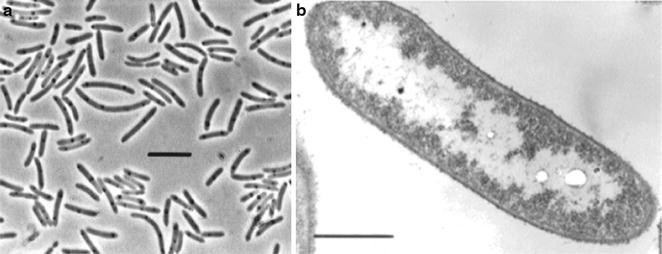
An image of Heliobacteriaceae.
- Chlorobi
- Aquificales (Some like it very hot! Samurai heart? iykyk)
- Nitrogen-Fixing Bacteria
These are an order of hyperthermophilic bacteria. They are also microaerophilic (they want only a small amount of oxygen!) and live in the sea near sea vents and in the land near hydrothermal systems.

This is an image of Aquifex, a genera of bacteria that is also called "water-maker" because they make water (duh) through the Knallgas Reaction. Their optimum temperature is 85 degrees Celcius.
These bacteria work with plants and are either symbiotic (lives inside the plant) or free-living (just in the soil). They turn atomospheric nitrogen into ammonia using nitrogenase. This is important because ammonia (NH4) is a form of nitrogen that plants and other microorganisms can use, along with nitrate (NO3-). These bacteria are super important. (pls review: Nitrogen cycle)
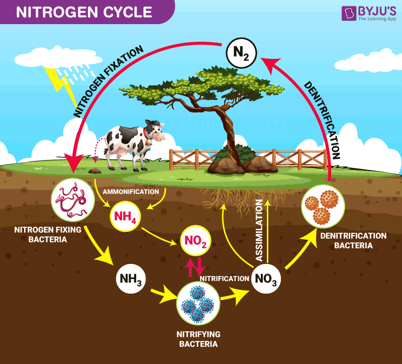
This is the Nitrogen cycle. Will expand on it later.
There is an interesting case...


This is Epulopiscium, a giant bacteria (0.6 mm; most bacteria are 0.2μm). It is able to be that big because it has many copies of of its genome throughout, and is thus an extreme case of polyploidy. It produces offspring inside its body (evolved from endospore!) and then the daughter cells later emerge.
Diversity of Archaea
Compared to bacteria, archaea is fewer. There are only 20 archaeal phyla recognized by 2016. By 2021, there are around 30 phyla. These are recognized using small unit ribosomal RNA as usual, which is like, the standard for phylogenetic analysis because it is conserved.
Archaea are usually extremophiles, but not always. Also: none of them are pathogenic to humans so far. This is likely because they have such extreme needs. Because of that, they are also extremely hard to culture in the lab.
So let's move on to groups of Archaea! In this lesson we will talk about three. Technically there's four, but they're onl the thermophiles, which is hyperthermophiles' less hot cousin.- Hyperthermophiles
- A lot of them are in the phyla Crenarchaeota.
- They are facultative heterotrophs. Which means: they NEED food from other organisms/organic matter to survive.
- They are found in volcanic areas where sulfur gases are emitted, hot springs, hydrothermal vents...
- Methanogens
- They are in phylum Euyarchaeota.
- They are the moderates of archaea because they live in moderate pH, temoerature, and salinity. The only special thing about them is that they produce methane, through methanogenesis. This is a process unique to archaea, no other domain does this!
- Methane is a major greenhouse gas, and 74% of methane in the atmosphere is produced by these bad boys...
- Most of them use H to reduce CO2, but others use formate, alcohol, and acetate too!
- Halophiles
- They are part of order Halobacteriales in phylum Euyarchaeota.
- They can live even in more than 150-200 g/L salt environments (salty!)
- They maintain high concentations to K+ and Cl- inside their cells to counteract all of the water inside them trying to go out to the salty world. If they're concentrated inside too, then they won't give in to this osmotic pressure.
- They have two pumps: bacteriorhodopsin, which pumps protons out of the cell (to maintain neutral pH) and halorhodopsin, which pump Cl- into the cell, which helps with that osmotic pressure. They are both activated with good ole light energy.

This is an image of a Pyrodictium. Pyrodictium occultum, which literally means "fire connection; hidden" can live in up to 110 deg C (boiling hot!) in shallow, hot volcano vents.

Pyrolobus fumarii, which means "fire lobe of the chimney" is another hyperthermophile than can live up to 113 deg C and lives in fumaroles (vents in the ocean that release mineral-rich, superheated water or black smokers.

Now this is an interesting one. Nanoarchaeum equitans, which literally means "very small archean rider" is the small orange cells in A and the smaller cell in B. The green and the bigger cells is Ignicoccus (which means fire sphere), specifically Ignicoccus hospitalis. These two have a special symbiotic relationship, and it almost looks as if N. equitans is 'riding the fire sphere' (kinky). The two of them live in submarine vents which can go from 70-90 deg C. N. equitans is also known as one of the smallest cellular organisms, with a genome size of 0.5 Mb. It is the only organism in phylum Nanoarcheota at this current time.
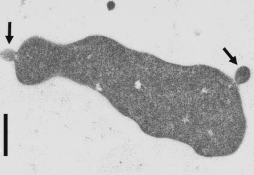
This is Ferroplasma acidiphilum, an iron-oxidizing, acid-loving archaean without a cell wall. They were cultured and desribed from a pyrite-leaching bioreactor. They are able to survive in such acidic places because their membranes do not allow a lot of protons (acid) to pass through, thus they manage to keep a relatively neutral internal pH. Also: because they accumulate iron oxide inside their cells, they are red!
Notable information:

Say hi to methanogens!
Notable information:
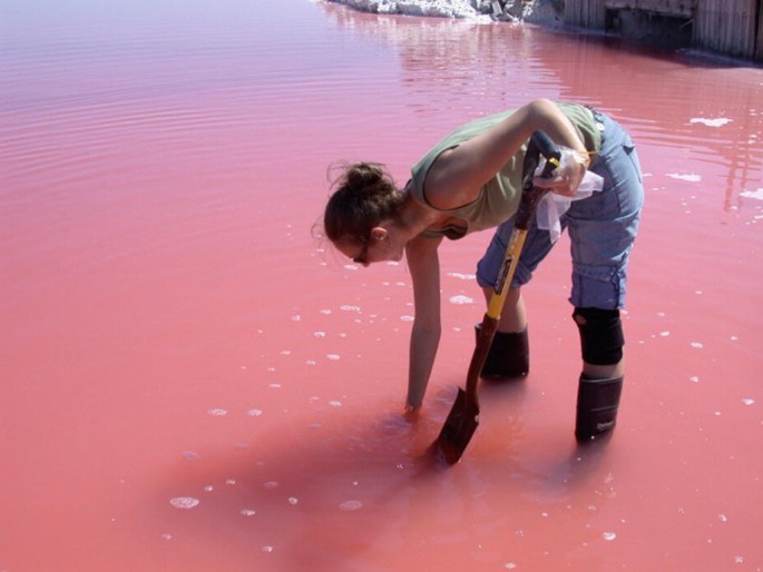
A hypersaline pool. It's pink! That's because of halophiles. Most of them have reddish/pinkish hue because they have carotenoids in the cell membranes.
Notable information:
REMEMBER!
Archaeal core housekeeping (the way they manage their insides) and their metabolic functions (the way they make their energy) is similar to bacteria, but their information-processing systems (DNA, RNA, and the central dogma) are more similar to eukaryotes.

CONGRATULATIONS! Another lesson (well, half a chapter, but it's such a long chapter isn't it?) DONE! Take a break and continue reading later.
2. Diversity of Microorganisms (Continued)
Date Finished: October XX, 2024
Now let's move on to Protist Diversity!
Diversity of Protists
So what's the deal with protists?
Protists can be unicellular and multicellular. They are widespread, they need water yadda yadda yadda. There are a lot of discourse on the taxonomy of protists but basically they usually fall on two groups:
- Protozoa - heterotrophic, motile and single-celled, basically the precursor to animals.
- Algae - photosynthetic and produces oxygen, basically the precursor to plants.
Protist Groups
- Diplomonads
- They contain TWO mitosomes (mitochondria which lost its primary function because they live in places without oxygen). That being said...
- They live in places without oxygen like animal intestines and urogenital tracts. They are either symbiotic or parasitic.
- They do fermentation and anaerobic metabolism... (Duh, no oxygen?)
- Parabasalids
- They have hydrogenosomes (like mitochondria but produce hygrogen gas)
- They, like Diplomonads, live in anoxic habitats as symbiotes or parasites, and also does fermentation and anaerobic metabolism.
- They typically only have one nucelus, unlike diplomonads.
- Euglenozoans
- They are unicellular and have a flagellar crystalline rod. This is a fancy way of saying that their flagella has crystalline proteins.
- They often have two flagella, but not always.
- Alveolates
- They have sacs in their cytoplasmic membrane called and alveoli (like in our lungs!), though this alveoli is used for structural support rather than gas exchange. They are not filled with air, but rather fluid or other substances.
- Stramenophiles
- They typically have two types of flagella. One is smooth and the other is covered with hair-like structures. This is where the name comes from, as "stramen" means hair-like structures.
- Amoebozoa
- They use pseudopodia, which is also known as "false feet".
- This group involves not only the famous amoeba but also the slime molds.

Diplomonads have flaggela, are unicellular and have two nuclei. In this photo, the Giardia lambia appears to be smiling. Those "eyes" are its nuclei. They cause common waterborne diarrheal disease.
Notable information:

This is Trichomonas vaginalis, which causes an STD (Trichosomiasis) in humans.
Notable information:

This is a group under Euglenozoans called the kinetoplastids. Yes, the're those dark pink squiggles. If you ask me, they look a little bit like a deflated plastic bag. They have a kinetoplast, which is a mass of circular DNA inside a single large mitochondrion. They are pathogenic. An example is Trypanosoma bruceii.

These, on the other hand, are Euglenids. They're the kinetoplastids' nonpathogenic sisters. They are phototrophicb (which is why they're green!), lives in water, and feed on bacterial cells. An example, of course, is the famous Euglena.<
Notable information:

One of the groups under this one are the ciliates. They use cilia for motility and food. They have two nuclei: the macronucleus (used for feeding) and the micronucleus (used for sexual purposes). A famous ciliate is our friend paramecium, shown above.
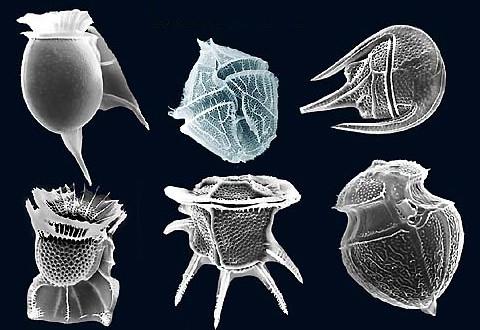
Another group is the dinoflagellates. They are phototrophs and has flagella. They love warm and polluted coastal waters, and that is why they are often the reason for red tide

A red tide. The red of the ocean is due to the carotenoid pigments of Gonyaulax cells. It paralyzes shellfish due to neurotoxins, and those toxins are harmful for the people that eat them.

These are apicomplexans. Notable members include Plasmodium (malaria), Toxoplasma (toxoplasmosis) and Eimeria (coccidiosis). They become sporozoites, which is their motile, infective stage. They then move to through the bloodstream and find their way to the liver. They also contain apicoplasts, which is like a chloroplast that got tired with life and decided to degenerate.
Notable information:

These are Diatoms. They are unicellular and phototrophic. They are also the planktonic phytoplankton, which is a fancy way to say they drift along the flow of water and are photosynthetic. They also have a frustule, which is a cell wall made of silica. It shows pinnate and radial symmetry and are really quite pretty.

These are Oomycetes. They look a lot like fungi (thus they are called egg fungi, but no they are not! An example seen here is Phytophtora infestans, which is a plant pathogen that causes blight disease in potatoes.
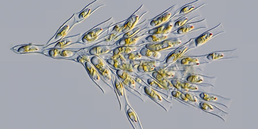
These are chrysophytes, which literally means "golden plant". They are unicellular, but they clump up and form colonies and thats why they look big to the naked eye. They are found both in marine and salt water and their pigment is duee to fucoxanthin carotenoid.

These are brown algae. Which supposedly is under stramenophiles too... I'm going to have to ask a confirmation from my prof because there seemed to be a mix-up about this. Will update later.
Notable information:
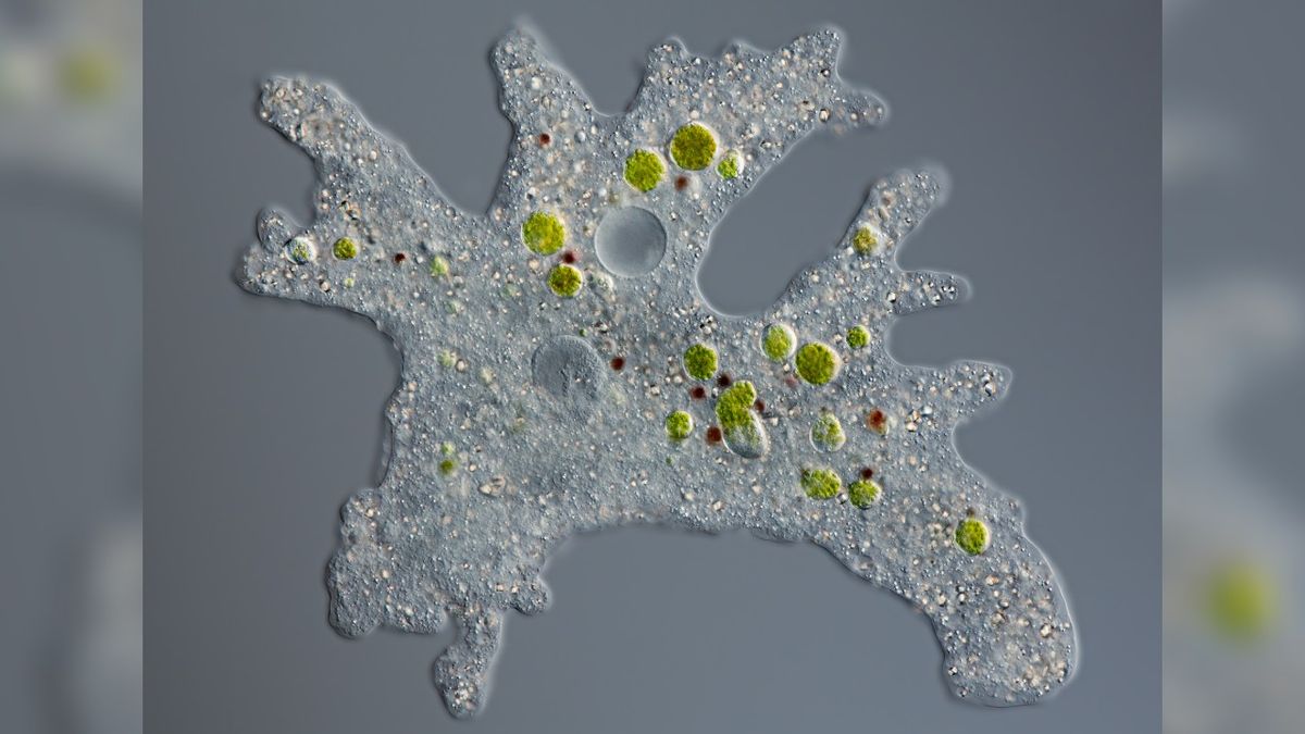
This is the famous Amoeba, which is part of the group Gymnamoebas. I guess they go to the gym or something.

This is Entomoeba histolytica, which is a part of the group, you guessed it, Entamoeba. They cause Amebiasis aka amebic dysentery which leads to holes in the intestines causes blood diarrhea. They are transferred from fecal contamination of water and food.
Notable information:
Diversity of Fungi
Here we go to what I personally find reall interesting: Fungi! Of course these includes our molds, yeasts, and mushrooms. Fungi can be unicellular or multicellular. They are non-vascular, which means that they do not have the vascular tissue that plants and animals do, and they are also obligate chemoheterotrophs, which means absolutely none of them are photosynthetic.
Fungi thrive from relationships with other organisms. In the first place, they eat dead organic organisms, but that's not where it ends. They also gave parasitic or mutualistic associations with insects and plants. Fungi helps plants absorb more nutrients from the soil. And have you never read a horror book about parasite fungus taking over our bodies and hijacking our brain?
Their cell walls contain chitin, which keeps it rigid, though some fungi also has cellulose, which you often find in plants. Their cell membrane contain ergosterol, which I guess is cholesterol's less famous and more pretentious cousin. Their spores can be produced sexually or asexually, which we will expand upon on the next part of the lesson.
[DBC] Developmental Biology Lecture
1. Introduction to Developmental Biology
Date Finished: October 15, 2024
Developmental Biology is the study of an organism's life cycle, from embryo to adult.
Historical Figures in Developmental Biology
| Hippocrates | Applied scientific approach to embryonic development (Scientific Method!) Explained development using heat, wetness and solidification. He believed that heat and wetness are important for the embryo, and that the liquid embryo "solidifies" into its proper shape |
|---|---|
| Aristotle | First to study embryos of different organisms Thought that male contibutes semen (because I guess semen is an obvious component of a baby during those times) and females contribute a wholeass embryo he called catamenia |
| Galen | Thought that embryos use umbilical cord to breathe (snorkeling baby??) Described the allantois and amnion, two of the four extraembryonic membranes, and placenta, which is connected to the umbilical cord. |

Those are the ancient dudes related to Developmental Biology. Now meditate and remember them before we move on. Have some kitty.
| Leonardo Da Vinci | Dissected the human fetus (he a lil bit freaky...) Measured development with NUMBERS First to give evidence that embryos change in weight, size and shape (Wild that this was not common knowledge in the past...) |
|---|---|
| William Harvey | Studied eggs! Observed chick embryo's circulatory system Found out that the white spot in the egg is where the embryo arises Believed that amniotic fluid was absorbed into the blood of the embryo |
| Marcelo Malphigi | First microscopic account of chick development Somehow concluded that there's a mini chick inside the egg (bro??) Because of that, Preformationism grew. |
What is Preformationism?
Preformation Theory is the idea that the embryo is already fully formed in the egg It's just small, and it just grows in size over time. The people who believed this were called ovists
On the other hand...
The men can't handle not being the center of attention (I kid, I kid) so they made a counterpart of Homunculus Theory, which posits that the mini embryo is in the head of every sperm instead. The people who believed this were called spermists.
Imagine if there truly was a million mini babies in each man's sperm...
| Caspar Wolff | Demonstrated that the chick intestine comes from tissue on the embryo's surface Proposed Theory of Epigenesis, which states that new structures arise through many stages (against Preformation, I suppose...) |
|---|---|
| Johann Wolfgang von Goethe | Studied angiosperms and believed there is a Bauplan (fundamental organization) in their diversity Realized that flowers are just modified leaves |
| Theodor Schwann and Matthias Jacob Schleiden | Proposed the Cell Theory |
:max_bytes(150000):strip_icc()/TC_373300-cell-theory-5ac78460ff1b78003704db92.png)
Remember Cell Theory? 1-3 is the classic theory, while 4-6 is the added modern version. This is one of the basis of biology.
| August Weismann | Proposed Germplasm Theory, which states that the body is divided into somatoplasm and germplasm. Basically somatic cells and germ cells (gametes) |
|---|---|
| Wilhelm Roux | Proposed Mosaic Theory |
| Han Driesch | Proposed Regulative Theory |
Mosaic Theory vs Regulative Theory

A shows an embyo with a development that is not disturbed at the far left. In the Mosaic Model, if the part of the embryo that is meant to be the head is disturbed, then the baby won't grow a proper head. In the Regulative Model, the cells would just communicate with each other and can shift around and make up for the damaged/disturbed cells until the baby is fully formed, well and not disfigured.
B shows Regulative Theory. Even if the cells shfit around, a functional baby is born.
So which is true?
Both! It depends on the organism and their stage of development. Some organisms of stage of developments show a mosaic pattern (like sea urchins), while some shows regulative pattern (like mammals and yes, including humans).
| Hans Spemann and Hilde Mangold | Proposed the idea of induction, which is connected to Regulative Theory. It is the idea that certain regions in the embryo can change the behavior of nearby cells. They can communicate and have teamwork to form the bab |
|---|

The Spemann-Mangold Organizer Experiment. Here, they took two newt embryos at the gastrula stage (where the cells start to differentiate) and grabbed a part now called the Spemann organizer from one embryo and transplated it to another. The organizer pushed nearby cells to create an entirely new conjoined body, which proved the idea of induction.
Development of Genetics through Developmental Biology
| Thomas Hunt Morgan | Established Chromosomal Basis of Heredity, which is a foundation of Modern Genetics |
|---|---|
| George Redei | Started the use of Arabidopsis thaliana as a model for dicot plant development studies |

Thomas Hunt Morgan's famous fruit fly experiment. Here, he took fruit flies with white eyes and red eyes. Through breeding, he figured out that that specific eye color is a recessive sex-linked trait which is linked to the X chromosome.
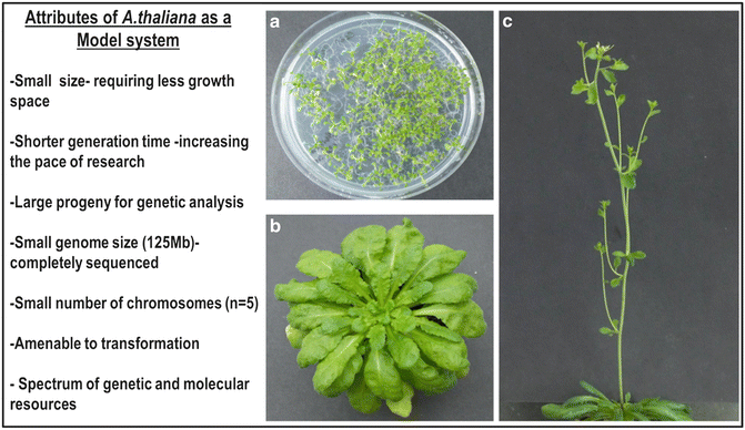
Arabidopsis thaliana, also known as thale cress: a model organism.
Basic Development Goes like This:
| Growth increase in size | Cell division increase in number | Differentiation Diversification of cell types | Pattern Formation organization or cells | Morphogenesis formation of shapes and structures |
|---|

CONGRATULATIONS! You finished a lesson. Good job!
2. The Plant: A Review
I imagine this lesson will be quick since this is just a review and I know a lot of this by heart already.
Plants! Our chlorophyllic neighbors. General characteristics include: multicellularity, being a primary terrestrial eukaryote, well-developed tissues, autotrophic by photosynthesis (duh), non-motile, and protect their embryo using a seed (which depends because some plants do not have seeds...). Vegetative structures includes roots, stems and leaves. Reproductive structures includes flowers, fruits and seeds.
Plansts have two organ systems. Everything under the soil(Root System and everything above the soil (Shoot system.
The Root System
Roots, obviously, help the plant get nutrients from the soil. It does so by creating root hairs, which greatly increase absroption because there's a lot of surface area. On the other hand, sometimes roots develop from the organs of the shoot system and go down to the ground. Sometimes this is for support. Sometimes I guess they just wanna hang around and look pretty, like in the Banyan Tree (I'm sure there is a very important adaptation-related reason. I just have no time to dig in it right now).
These are the root hairs. As you can see, the root hair is actually just one epidermis cell that extends beyond. The greater surface area allows for more room for nutrients to do their osmosis thing and get inside.
These are examples of adventitious roots from Zea mays (corn) and Ficus (Banyan Tree).
There are two types of roots: taproot, which is common in dicots, and fibrous, which is common for monocots.
The Shoot System
The stems help support the leaves, flowers and fruits and act as the highway through which water and food are transported (duh).
But here's the interesting thing! Sometimes, they can become metamorphosed stems, which is fancy way to say modifed stems. For adaptation, plants have modified their stems into many different things! They can be:
- Photosynthetic - They aren't, usually! When you see green stems, they can photosynthesize for sure.
- Tendrils - These are special stems for climbing. You know the one.
- Thorn - Surprise! Rose thorns? Stems.
- Bulb - underground, fleshy bud. Think onions. Onions may be underground but they are not roots. The roots grow under them remember? They are stems!
- Corm - round, perennial stem with papery leaves, my prof says... but I don't think I've ever seen one before. It's like a bulb... but it has no layers. Just... solid in there. Taro is apparently an example.
- Stolon aka runner, this one is a reproductive stem that grows abobve ground.
- Rhizome - this, on the other hand, is a reproductive stem that grows below ground.
- Tuber - enlarged tip of a rhizome. Think potatoes. They are not roots. They are stems. That's why potatoes have buds (or "eyes") that can sprout new baby potato plants.

So let's move on to the leaves.
[DBB] Developmental Biology Laboratory
1. Bryophytes and Pteridophytes
Date Finished: October 24, 2024
This lab activity is about non-vascular (Bryophytes) and lower vascular (Pteridophytes) and their reproductive structures. It's quite interesting actually. So strap in.
Laboratory Procedure
A/B. Bryophytes/ Pteridophytes
- Look around the campus and collect pictures of mosses, liverworts, and hornworts.
- Label the shit out of these pictures.
- Label the shit out of the pictures given by the prof.
- Same procedure but with ferns.
- Profit?
C. Spore Dissection
- Basically, gather up em sori from ferns.
- Look at the spores under dissecting and light microscope.
- Take a picture and label the shit out of those pictures.
- Profit.
So what are the concepts?
As earlier stated, these are non-vascular and lower vascular plants. But what does it mean?
Non-vascular means that these plants do not have vascular tissues, also known as the xylem and phloem. These are basically tubes that run along the inside of the plant from root to leaves that transport water (xylem) and food (phloem) to different parts of the plant. Not having this is characteristic of those plants which are earlier in the evolutionary tree and are much closer to the algal anscestors. They didn't manage to develop it yet, and thus has adapted to living without.
Lower vascular plants, on the other hand, mean that they do have these vascular tissues, just that they are still fairly simple and are not as advanced as the gymnosperms and angiosperms, which we will be learning about in the next activity.

This is the Plant Phylogenetic tree. As you can see, the Bryophytes and Seedless Vascular plants (which included lycophytes aka club mosses and pteridophytes aka ferns) is closer to the anscestor of all plants. There is something much closer, called the Charophytes. They are not true plants, as they are a group of green algae and are considered the closest relative to land plants.
Reproductive Cycle
All plants undergo in what we call Alternation of generations. This means that the reproductive cycle goes through a gametophyte generation and a sporophyte generation and to a gametophyte generation over and over again in a circle. In the case of byrophytes, the dominant generation (the generation that you can most easily see and identify as: ha! That's the plant!) is the gametophyte generation.
Gametophte generation are haploid while the sporophyte generation are diploid. This means that if the organism has 46 chromosomes, a sporophyte would have 46 chromosomes while a gametophyte will only have half, which is 23.
Confused? It is confusing, a little bit! But let's break it down through pictures!
In the simplest terms, the alternation happens like this:
| The gametophyte produces the gametes through mitosis. |
|---|
| Two types of gametes (male and female) fuse together to become a diploid zygote. This process is called fertilization. |
| The zygote divides and becomes a sporophyte through mitosis. |
| The sporophyte produces spores through meiosis, cutting the chromosomes in half. |
| A single spore becomes a gametophyte (male or female or both) through mitosis. |
| The cycle continues. |
How does this look like in practice?
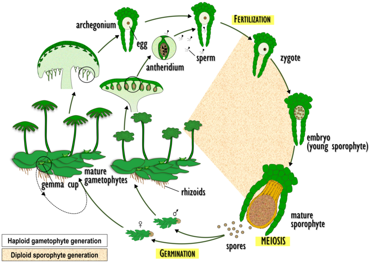
The alternation of generations in liverworts.

The alternation of generations in mosses.

The alternation of generations in hornworts.
Don't worry if it all looks very technical! We will tackle each group of bryophytes one by one.
The dominant generation of ferns, on the other hand, is the sporophyte generation. You will see that this is actually the more common dominant generation to higher vascular plants.

The alternation of generations in ferns.
Bryophytes
As promised: let's talk about each group one by one. There are three groups of bryophytes, and they are all cute and lovely.I had so much fun looking for them all around campus.
- Liverworts
- Thallus - the body of the liverwort. That flat leaf-like thing.
- Rhizoid - the "roots" of the liverwort. It provides anchorage.
- Archegoniophore - the structure that holds the archegonium (egg-producing organs).
- Antheridiophore - the structure that holds the antheridium (sperm-producing organs).
- Gemmae - a special structure unique to liverworts that allow them to reproduce asexually. These are small "cups" on the thallus that when hit by water can launch mini clones of the liverworts farther along where it won't compete with the "parent".
- Midrib - like in leaves, it's the middle of the thallus.
- Capsule - the part of the sporophyte where the spores are developed. Once mature, it explodes spores everywhere. These spores eventually become new gametophytes
- Seta - the part of the sporophyte that supports the capsule and keeps it attached in the archegonium.
- Antheridia - the sperm-producing part of the plant.
- Archegonia - the egg producing part of the plant (where the sporophyte hangs off of!)
- Stalk - aka peduncle. This is the... well, stalk part of the antheridiophore or archegoniophore.
- Neck - the part of an archegonia leading to the egg where the sperm enters.
- Egg - the female gametes, obviously.
- Ventor- the cavity that houses the archegonia.
- Mosses
- Antheridia
- Archegonia
- Stalk
- Neck
- Egg
- Vector
- Antheridia
- Archegonia
- Stalk
- Neck
- Egg
- Vector
- Hornworts
- Antheridia
- Archegonia
- Stalk
- Neck
- Egg
- Vector

This is the parts of a liverwort.
Notable parts:

This is what a gemmae actually looks like in real life.

This is an archegoniophore up close. The yellow stuff is the sporophyte. They look like little palms with fruits.
A little bit closer...

Antheridiophore cross-section under the microscope, labeled.

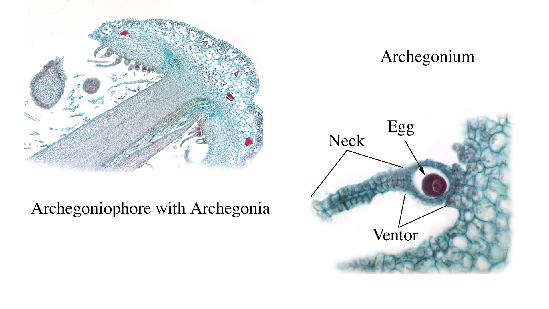
Archegoniophore cross-section under the microscope, labeled.

Liverwort sporophyte cross-section under the microscope, labeled.
Notable parts:
Notable parts:
A little bit closer...
Notable parts:
Notable parts: